

News
IIS La Fe Nursing Professionals Lead a Study on the Use of Fluorescence Imaging to Detect Bacteria in Chronic Wounds
A study led by nursing professionals from the Dermatology and Tissue Regeneration Research Group at the Health Research Institute La Fe (IIS La Fe) has demonstrated the usefulness of fluorescence imaging for the early detection of clinically significant bacterial loads in hard-to-heal wounds that do not yet show obvious signs of infection.
The study, conducted in collaboration with the Dermatology Department of the Hospital Universitari i Politècnic La Fe, shows that this technology provides immediate information on the presence of bacteria during a clinical consultation. The findings have been published in the scientific journal Journal of Wound Management.
The research included patients with wounds that had been present for more than six weeks and showed no obvious clinical signs of infection. The team used a portable fluorescence imaging device to visualise the presence of certain bacteria directly on the wound bed. The findings were subsequently compared with the results of conventional microbiological cultures.
The results showed a high level of agreement between the two techniques. Fluorescence imaging achieved a sensitivity of 90.9% and an overall diagnostic accuracy of 85% in identifying a clinically significant bacterial load.
These findings support the potential of this tool as a complement to clinical assessment and as an aid in making decisions about wound treatment.
Real-Time Microbiological Information
Fluorescence imaging is a non-invasive technique that enables the real-time detection of signals associated with a high bacterial load. Its use may be particularly relevant in wounds that do not display the classic signs of infection but whose clinical progression raises suspicion of significant bacterial colonisation.
The information obtained through this technology can help clinicians select the most appropriate areas for microbiological sampling. It can also support wound bed preparation and the planning of local treatment.
Its use as a complementary tool could also contribute to more personalised care and more rational use of antimicrobials. However, the technique does not replace clinical assessment or conventional microbiological procedures when these are indicated.
Research Applied to Improving Patient Care
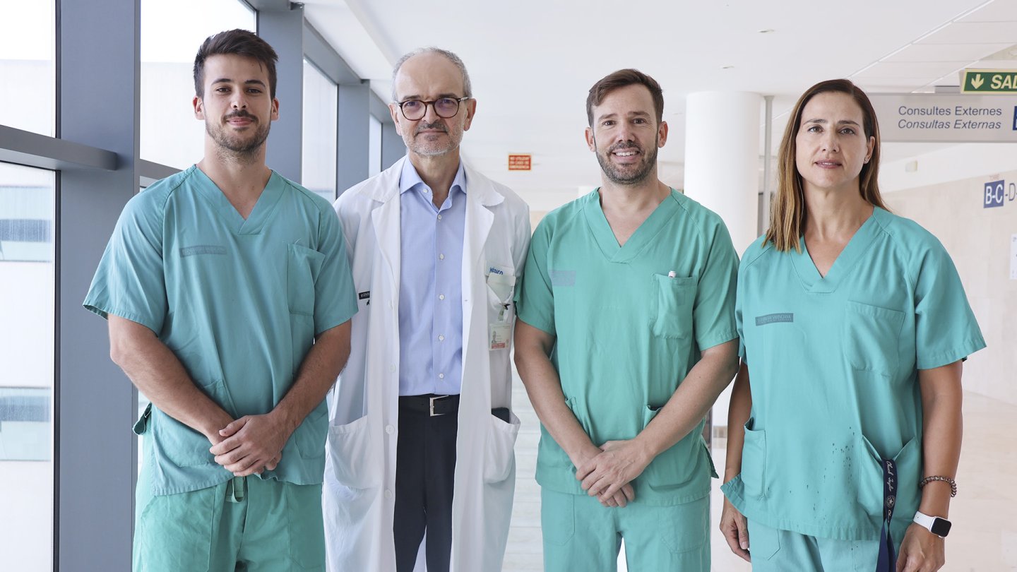
The study was led by nurse researchers Miguel Ángel Parada-Sanchis and Víctor Gadea-López, members of the Dermatology and Tissue Regeneration Research Group at IIS La Fe and the Dermatology Department of the Hospital Universitari i Politècnic La Fe, together with dermatologist Rafael Botella-Estrada.
The study highlights the contribution of nursing professionals to the generation of scientific knowledge and the evaluation of new technologies applicable to patient care. Their participation in multidisciplinary research groups enables projects aimed at improving the quality, safety and efficiency of healthcare.
The research also underscores the value of collaboration between nursing and medical professionals in translating scientific evidence into clinical practice and exploring new solutions to complex problems, such as chronic or hard-to-heal wounds.
This publication reflects IIS La Fe's commitment to translational, multidisciplinary research focused on patients' needs.
The evaluation of non-invasive diagnostic tools available at the point of care may contribute to the development of more precise and efficient healthcare models. In this context, IIS La Fe promotes the participation and leadership of nursing professionals in research projects with direct applications in clinical practice and in improving health outcomes.
Parada-Sanchis MA, Gadea-López V, Botella-Estrada R. Detection of subclinical infection using fluorescence imaging in hard-to-heal wounds. Journal of Wound Management. 2026;27(3). DOI: 10.35279/jowm2026.27.03.03.
Share new


